Taste and Smell

35

Learning Objectives

Know where taste information first reaches the brain.

Know where the nucleus of the solitary tract is and what role it plays in relaying taste information to the brain.

Taste buds are formed by groupings of taste receptor cells. Receptor cells protrude into the central pore of the taste bud. Chemical changes within sensory cells, from the taste molecules binding onto the taste receptors, will result in neural impulses that transfer to the brain through other nerves. Where these neural impulses transfer to will depend on where the receptor is located, such as the posterior two thirds of the tongue. Taste information is transmitted to the medulla, thalamus, limbic system, and to the gustatory cortex, which is tucked underneath the overlap between the frontal and temporal lobesĀ (Maffei, Haley, & Fontanini, 2012; Roper, 2013).
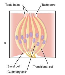
 

Each taste bud consists of a pore, which is an indentation in the surface of the tongue. There taste hairs reside and lead to three different types of cells: basal cells, gustatory cells, and transitional cells.
Figure 4.7.1. Taste Buds. Taste buds are composed of multiple taste receptor cells. Unlike olfactory neurons, taste cells do not have axons. Taste buds contain gustatory cells, basal cells, and transitional cells. The gustatory cells contain hair like cells called microvilli that extend through the taste pore in the taste buds. These taste cells transmit information by using secondary afferent neurons to make synaptic contact with taste cells to transmit information to the thalamus. (Credit: Cheryl Olman, own photo. CC-BY-4.0)


Due to the fact that taste cells have no axons, secondary afferent neurons with cell bodies in the nucleus of the solitary tract, which is located in the medulla of the brain stem, make synaptic contact with taste cells. Depending on where these cells and neurons come from, they will terminate in specific parts of the solitary nucleus. After termination at the solitary nucleus, the new information will relay data to the thalamus. In addition, information from the thalamus is transported to the frontal operculum and insular cortex, which is the primary taste area of the brain.
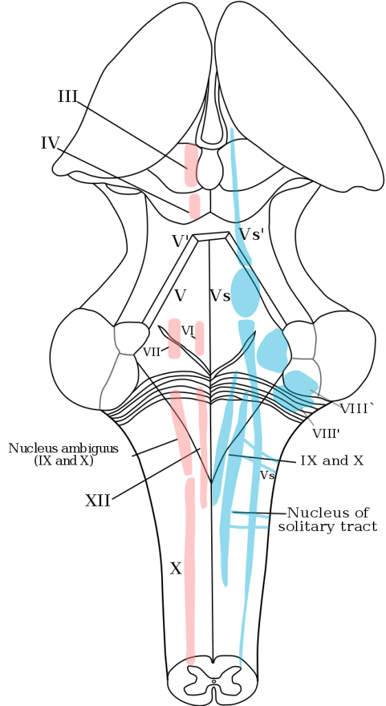
 

Depicts a posterior view of the brain with the thalamus dissected out and the cerebellum removed. The 4th ventricle is splayed open in the center of the image. Major motor (red) and sensory (blue) nuclei are depicted.
Fig.4.7.2. Taste Data. Location of the nucleus of the solitary tract and insular cortex (primary gustatory cortex). The NST is very low in the brainstem. It is an important site for the modification of taste information either by blood glucose (which inhibits responses) or by feedback from cortical regions like the orbitofrontal cortex. From the NST, taste information continues to the thalamus. (Credit: Mcstrother. Provided by: Wikipedia. License: Public Domain)


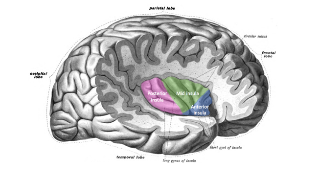
This image divides the insula into its anterior, mid, and posterior regions, with each being denoted by different colors.
Fig.4.7.3. The insular cortex is tucked behind the frontal and temporal cortexes, which is cut away in this illustration. The insular cortex is involved in more than just our sense of taste; it receives input from all over the brain with information about how our body is doing. The dorsal (top) part of the mid-insula is where taste information from the thalamus is represented in the brain. (Credit: Schappelle. Provided by: Wikimedia Commons. License: CC-BY SA 4.0)


CC LICENSED CONTENT, SHARED PREVIOUSLY
OpenStax, Psychology Chapter 5.5 The Other Senses
Provided by: Rice University.
Access for free at https://cnx.org/contents/Sr8Ev5Og@12.2:Nw9FOKLs@13/5-5-The-Other-Senses
License: CC-BY 4.0
Adapted by: Maria XiongCheryl Olman PSY 3031 Detailed Outline
Provided by: University of Minnesota
Download for free at http://vision.psych.umn.edu/users/caolman/courses/PSY3031/
License of original source: CC Attribution 4.0
Adapted by: Maria Xiong

License

Icon for the Creative Commons Attribution 4.0 International License

Introduction to Sensation and Perception Copyright © 2022 by Students of PSY 3031 and Edited by Dr. Cheryl Olman is licensed under a Creative Commons Attribution 4.0 International License, except where otherwise noted.

Share This Book