Visual Development and Object Recognition
113
Learning Objectives
Know where FFA (fusiform face area) and VWFA (visual word form area) are located.
Know the functions of these two areas.
Know which pathways (dorsal or ventral) the two areas belong to.
While we memorize a few regions as examples of specialized functions, it is more important to remember that every visual stimulus is processed by every visual region. Neurons selective to particular aspects of an image will be most responsive. Visual experience is the sum of all of these responses, and these regions provide simultaneous, multi-dimensional representations of our complex visual world.
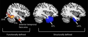
The FFA is located in the ventral stream on the ventral surface of the temporal lobe on the lateral side of the fusiform gyrus. This area of the brain has been specialized for facial recognition.
The visual word form area (VWFA) is a functional region of the left fusiform gyrus and surrounding cortex (right-hand side being part of the fusiform face area) that is hypothesized to be involved in identifying words and letters from lower-level shape images, prior to association with phonology or semantics.[1][2] Because the alphabet is relatively new in human evolution, it is unlikely that this region developed as a result of selection pressures related to word recognition per se; however, this region may be highly specialized for certain types of shapes that occur naturally in the environment and are therefore likely to surface within written language.[1] This area is also apart of the ventral stream.
Similar to auditory stimuli, more dorsal regions of the posterior half of the brain (parietal cortex) are involved in processing information about location (Where-and-How pathway); more ventral (temporal) regions are involved in recognizing objects (What pathway). Magnocellular information (colorblind neurons responding to large, fast things) tends to serve up the Where-and-How pathway. Parvocellular information (color-sensitive neurons responding to small, slow things) tends to head down the What pathway.
Again, a car zooming by is represented in the dorsal and ventral visual streams since we need information about both identity and location. Additionally, this information needs to be integrated. There is a massive white matter bundle (fasciculus) connecting dorsal and ventral regions. This paper by Jason Yeatman et al. on the history of the vertical occipital fasciculus is great:
Yeatman JD, Weiner KS, Pestilli F, Rokem A, Mezer A, Wandell BA (2014). The vertical occipital fasciculus: a century of controversy resolved by in vivo measurements. Proc Natl Acad Sci U S A. 111(48):E5214-23.
Cheryl Olman PSY 3031 Detailed Outline
Provided by: University of Minnesota
Download for free at http://vision.psych.umn.edu/users/caolman/courses/PSY3031/
License of original source: CC Attribution 4.0Wikipedia, Fusiform Face Area
URL:https://en.wikipedia.org/wiki/Fusiform_face_area#:~:text=Anatomical%20terminology,gyrus%20(Brodmann%20area%2037).
License: CC BY SA 3.0Wikipedia, Visual Word Form Area
URL: https://en.wikipedia.org/wiki/Visual_word_form_area
License: CC BY SA 3.0
