Vision Loss and V1
95
Learning Objectives
Know how the V1 neurons are organized according to eye of origin.
Know how the V1 neurons are organized according to their orientation preferences (depth vs surface).
Know what V1 hypercolumns are.
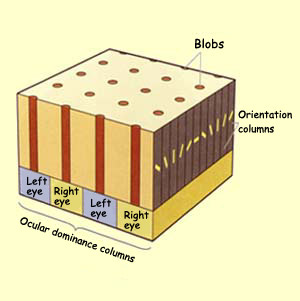
Columnar organization is an important property of primary sensory and motor cortices. The gray matter has 2 important directions: across the surface and through the depth.
- If you sample neural responses as you move through the depth, they stay approximately the same. This means that as you move down through cortex, you find neurons with the same orientation preferences.
- If you sample neural responses as you move across the surface, they change. For V1, this means that as you move across the cortex, you find neurons with different orientation preferences.
Left and right eye inputs are segregated into ocular dominance columns. This segregation is strongest in the input layer (4), so when we look for ODCs, we look in the middle of the cortex. A cluster of orientation columns is called a pinwheel—there’s an orientation pinwheel for each eye. Some V1 neurons are color-blind; some are color-selective. Color-selective neurons live in blobs (regions of cortex that stain dark when you stain for cytochrome oxidase, because they’re metabolically rich); there’s a blob for each pinwheel.
A cortical column is a group of neurons in the cortex of the brain that can be successively penetrated by a probe inserted perpendicularly to the cortical surface, and which have nearly identical receptive fields. Neurons within a minicolumn (microcolumn) encode similar features, whereas a hypercolumn “denotes a unit containing a full set of values for any given set of receptive field parameters.”
Cheryl Olman PSY 3031 Detailed Outline
Provided by: University of Minnesota
Download for free at http://vision.psych.umn.edu/users/caolman/courses/PSY3031/
License of original source: CC Attribution 4.0Wikipedia, Cortical column
URL: https://en.wikipedia.org/wiki/Cortical_column
License: CC BY-SA 3.0
