Light and Eyeballs
75
Learning Objectives
Know the idea of eccentricity in degrees, and how big our fovea and macula are.
Be able to describe the density of cones and rods from the fovea to the periphery.
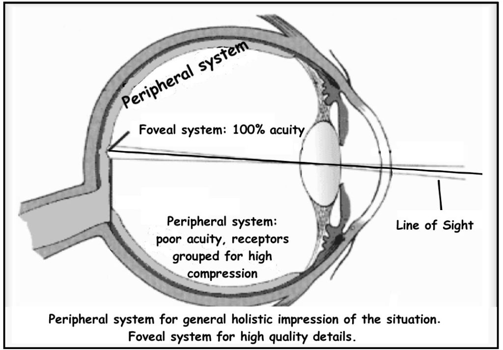
Know the visual acuity in our foveal vision and peripheral vision.
In a normal-sighted individual, the lens will focus images perfectly on a small indentation in the back of the eye known as the fovea, which is part of the retina, the light-sensitive lining of the eye. The fovea contains densely packed specialized photoreceptor cells. These photoreceptor cells, known as cones, are light-detecting cells. The cones are specialized types of photoreceptors that work best in bright light conditions. Cones are very sensitive to acute detail and provide tremendous spatial resolution. They also are directly involved in our ability to perceive color.
Although the macula comprises only 4% of the retinal area and 10% of the entire visual field, it is responsible for the majority of useful photopic vision. The fovea lies at the center of the macula and is approximately 2mm in diameter. The fovea is the central 1 degree of the visual field; the macula is the central 5 degrees of visual angle. The fovea contains the highest density of cone photoreceptor cells and is the only region of the retina where 20/20 vision is attainable. We have high acuity (ability to see fine detail) in the fovea because there is little convergence—basically one photoreceptor for every output ganglion cell. Acuity is also improved by high light levels (smaller pupil, ideally 2-5mm), longer exposure times, and appropriate focusing of the image by cornea and lens.
While cones are concentrated in the fovea where images tend to be focused, rods (another type of photoreceptor) are located throughout the remainder of the retina. Rods are specialized photoreceptors that work well in low light conditions, and while they lack the spatial resolution and color function of the cones, they are involved in our vision in dimly lit environments as well as in our perception of movement on the periphery of our visual field.
The peripheral retina is dominated by rods, but also contains cones. We have low acuity in the periphery because there is strong convergence: many rods map to a single output ganglion cell (since rods are more sensitive to light than cones). Convergence, in which each ganglion cell pools responses from multiple photoreceptors, increases sensitivity but decreases acuity. There is lots of convergence for rods and much less convergence for cones.
CC LICENSED CONTENT, SHARED PREVIOUSLY
Webvision: The Organization of the Retina and Visual System, Age-related macular degeneration (AMD)
Authored by: Gregory S. Hageman, Karen Gaehrs, Lincoln V. Johnson and Don Anderson
URL: https://webvision.med.utah.edu/book/part-xii-cell-biology-of-retinal-degenerations/age-related-macular-degeneration-amd/
License: CC BY-NC
Adapted by: Kori Skrypek
OpenStax, Psychology Chapter 5.3 Vision
Provided by: Rice University.
Download for free at http://cnx.org/contents/4abf04bf-93a0-45c3-9cbc-2cefd46e68cc@5.103.
License: CC-BY 4.0
Adapted by: Kori Skrypek
Cheryl Olman PSY 3031 Detailed Outline
Provided by: University of Minnesota
Download for free at http://vision.psych.umn.edu/users/caolman/courses/PSY3031/
License of original source: CC Attribution 4.0
Adapted by: Kori Skrypek
