Taste and Smell
29
Learning Objectives
Be able to locate the olfactory epithelium, the olfactory bulb, and the olfactory cortex.
Be able to describe an olfactory neuron and explain what a cilium is.
Understand that each olfactory neuron expresses only one type of receptor, and know approximately how many types of olfactory receptors there are.
Like taste, the sense of smell, or olfaction, is also responsive to chemical stimuli. The olfactory receptor neurons are located in a small region within the superior nasal cavity (see Figure 4.1.1 below). This region is referred to as the olfactory epithelium and contains bipolar sensory neurons. Each olfactory sensory neuron has dendrites (also called cilia) that extend from the apical surface of the epithelium into the mucus lining the cavity.
Cilia are small hair-like structures that can be found protruding from the olfactory receptor cells’ dendrites. The surface of these cilia is covered with olfactory receptors, also known as odorant receptors. An odorant will dissolve into the mucus of the olfactory epithelium and then bind to an olfactory receptor. Olfactory receptors can bind to a variety of odor molecules with varying affinities. The difference in affinities causes differences in activation patterns resulting in unique odorant profiles.
As airborne molecules are inhaled through the nose, they pass over the olfactory epithelial region and dissolve into the mucus. These odorant molecules bind to proteins that keep them dissolved in the mucus and help transport them to the olfactory dendrites. The odorant–protein complex binds to a receptor protein within the cell membrane of an olfactory dendrite. These receptors are G protein–coupled and will produce a graded membrane potential in the olfactory neurons.
The nasal epithelium, including the olfactory cells, can be harmed by airborne toxic chemicals. Therefore, the olfactory neurons are regularly replaced within the nasal epithelium, after which the axons of the new neurons must find their appropriate connections in the olfactory bulb. These new axons grow along the axons that are already in place in the cranial nerve.
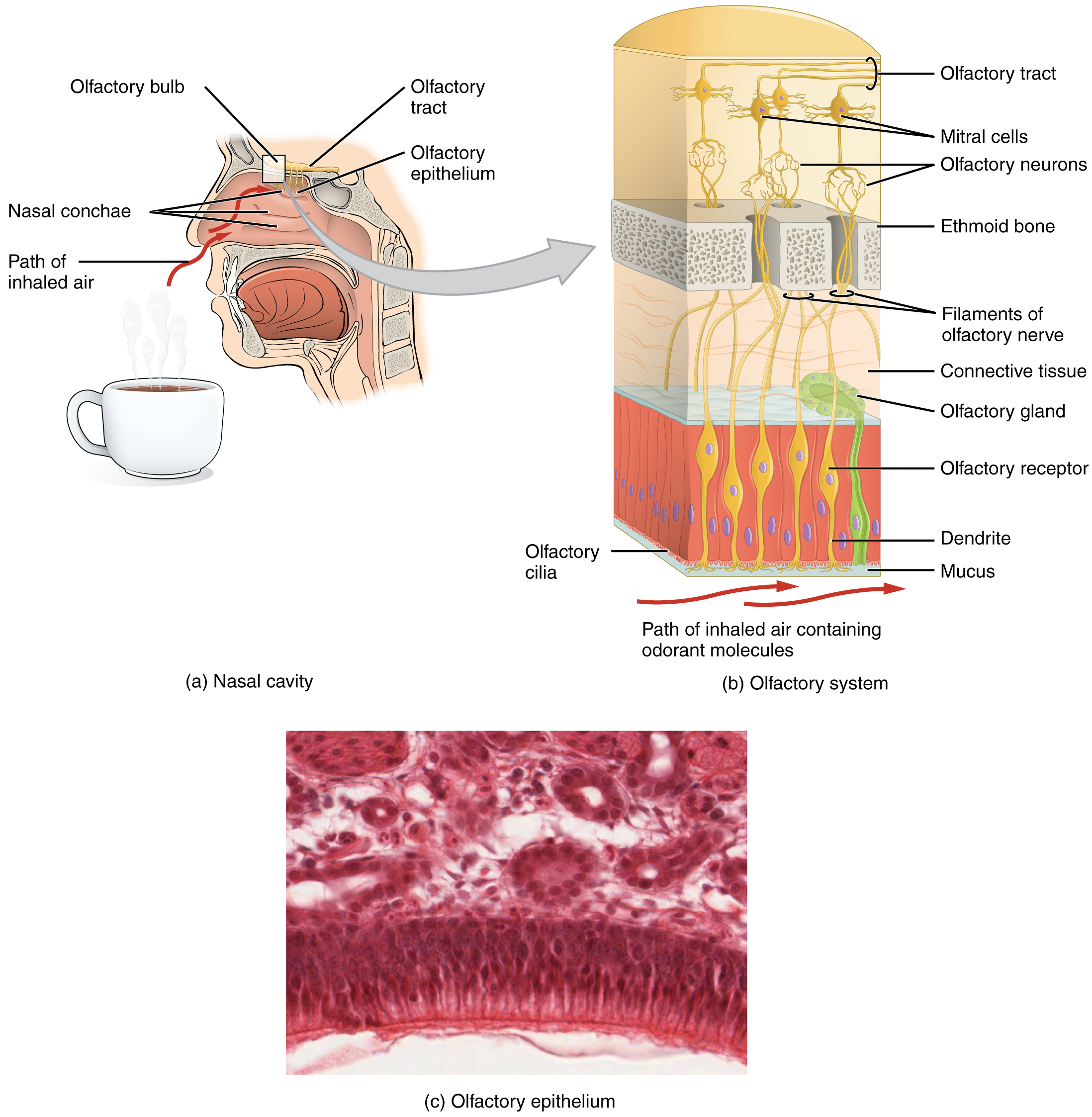
The axon of an olfactory neuron extends from the basal surface of the epithelium, through an olfactory foramen in the cribriform plate of the ethmoid bone, and into the brain (Fig.4.1.1). The first olfactory synapse in the brain occurs in a glomerulus in the olfactory bulb. A glomerulus is a cluster of synapses that collects information from olfactory neurons expressing a single type of receptor. There are about 350 different types of olfactory receptors, so there are about 350 different types of glomeruli spread throughout the olfactory bulb. Each receptor responds with different strengths to different kinds of molecules, so the activity level of a single type of olfactory receptor neuron (and therefore an individual glomerulus) does not predict what something will smell like. But the pattern of responses across the different glomeruli in the olfactory bulb provides a code from which you can decipher what something smells like to an individual.
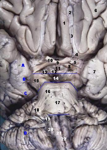
The group of axons called the olfactory tract connect to the olfactory bulb on the ventral surface of the frontal lobe. From there, the axons split to travel to several brain regions. Some travel to the cerebrum, specifically to the primary olfactory cortex that is located in piriform cortex (Fig.4.1.2) in the inferior and medial areas of the temporal lobe. Other axons from the olfactory bulb project to structures within the limbic system and hypothalamus, where smells become associated with long-term memory and emotional responses. This is how certain smells trigger emotional memories, such as the smell of food associated with one’s birthplace. Smell is the one sensory modality that does not synapse in the thalamus before connecting to the cerebral cortex. This intimate connection between the olfactory system and the cerebral cortex is one reason why smell can be a potent trigger of memories and emotion.
CC LICENSED CONTENT, SHARED PREVIOUSLY
OpenStax, Anatomy and Physiology Chapter 14.1 Sensory Perception
Provided by: Rice University.
Access for free at https://openstax.org/books/anatomy-and-physiology/pages/14-1-sensory-perception
License: CC-BY 4.0
Adapted by: Shawna Ratanpal
Wikipedia, Olfactory Receptor Neuron
Provided by: Wikipedia
URL: https://en.wikipedia.org/wiki/Olfactory_receptor_neuron
License: CC BY SA
