22.4 Oropharyngeal and Nasopharyngeal Suctioning Checklist & Sample Documentation
Open Resources for Nursing (Open RN)
Suctioning via the oropharyngeal (mouth) and nasopharyngeal (nasal) routes is performed to remove accumulated saliva, pulmonary secretions, blood, vomitus, and other foreign material from these areas that cannot be removed by the patient’s spontaneous cough or other less invasive procedures. Nasal and pharyngeal suctioning are performed in a wide variety of settings, including critical care units, emergency departments, inpatient acute care, skilled nursing facility care, home care, and outpatient/ambulatory care. Suctioning is indicated when the patient is unable to clear secretions and/or when there is audible or visible evidence of secretions in the large/central airways that persist in spite of the patient’s best cough effort. Need for suctioning is evidenced by one or more of the following:
- Visible secretions in the airway
- Chest auscultation of coarse, gurgling breath sounds, rhonchi, or diminished breath sounds
- Reported feeling of secretions in the chest
- Suspected aspiration of gastric or upper airway secretions
- Clinically apparent increased work of breathing
- Restlessness
- Unrelieved coughing[1]
In emergent situations, a provider order is not necessary for suctioning to maintain a patient’s airway. However, routine suctioning does require a provider order.
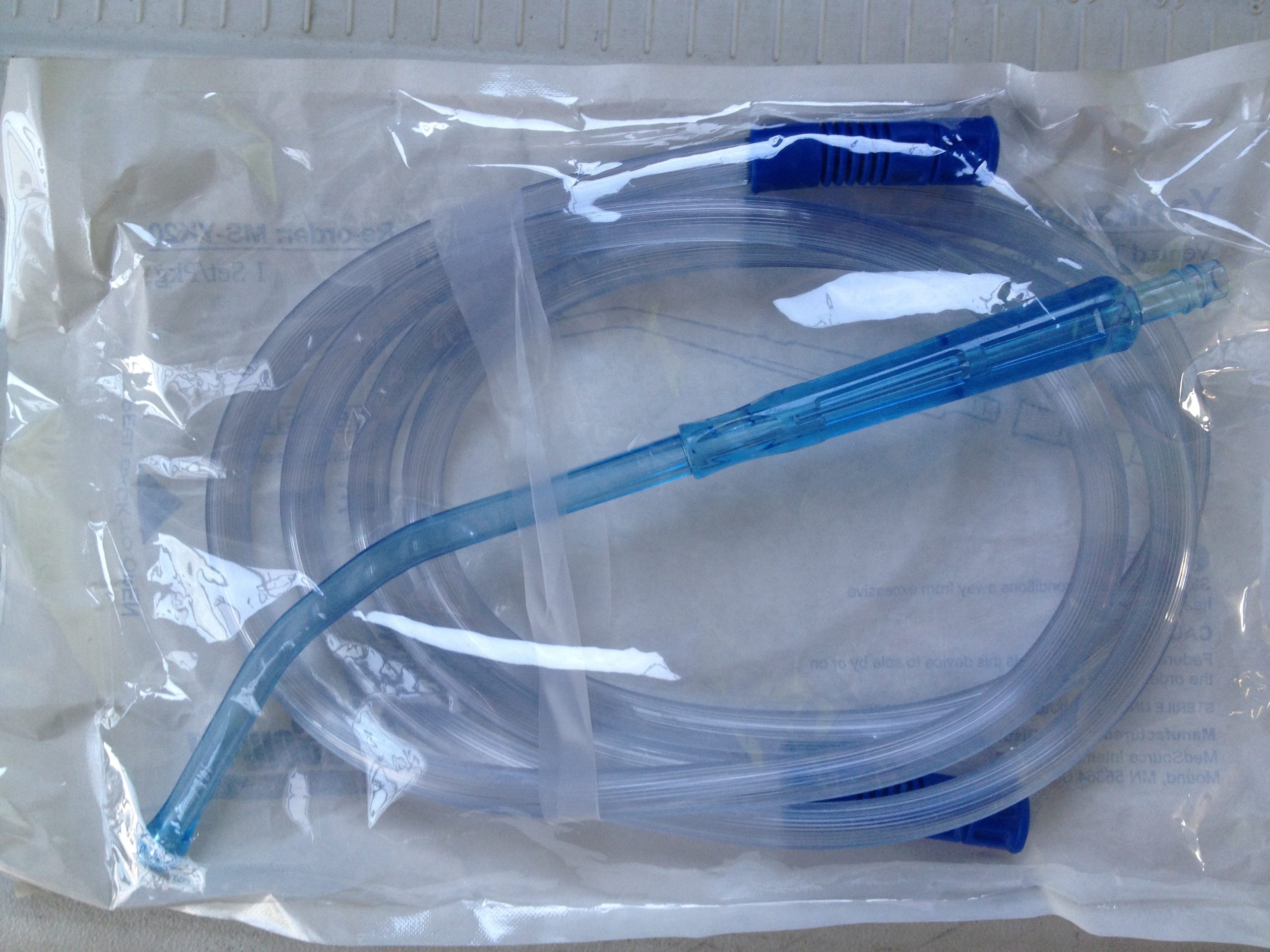
For oropharyngeal suctioning, a device called a Yankauer suction tip is typically used for suctioning mouth secretions. A Yankauer device is rigid and has several holes for suctioning secretions that are commonly thick and difficult for the patient to clear. See Figure 22.5[2] for an image of a Yankauer device. In many agencies, Yankauer suctioning can be delegated to trained assistive personnel if the patient is stable, but the nurse is responsible for assessing and documenting the patient’s respiratory status.

Nasopharyngeal suctioning removes secretions from the nasal cavity, pharynx, and throat by inserting a flexible, soft suction catheter through the nares. This type of suctioning is performed when oral suctioning with a Yankauer is ineffective. See Figure 22.6[3]for an image of a sterile suction catheter.
![“DSC_0210-150x150.jpg” by British Columbia Institute of Technology (BCIT) is licensed under CC BY 4.0. [/footnote] Access for free at https://opentextbc.ca/clinicalskills/chapter/5-7-oral-suctioning/ Photo of a sterile suction catheter being handled by a person wearing gloves](https://opentextbooks.uregina.ca/app/uploads/sites/160/2021/06/DSC_0210-scaled-1.jpg)
Extension tubing is used to attach the Yankauer or suction catheter device to a suction canister that is attached to wall suction or a portable suction source. The amount of suction is set to an appropriate pressure according to the patient’s age. See Figure 22.7[4] for an image of extension tubing attached to a suction canister that is connected to a wall suctioning source.
![“DSC_0206-e1437445438554.jpg” by by British Columbia Institute of Technology (BCIT) is licensed under CC BY 4.0. [/footnote]. Access for free at https://opentextbc.ca/clinicalskills/chapter/5-7-oral-suctioning/ Photo showing tubing attaching suction canister to wall suction source](https://opentextbooks.uregina.ca/app/uploads/sites/160/2021/06/DSC_0206-e1437445438554-scaled-1.jpg)
Follow agency policy regarding setting suction pressure. Pressure should not exceed 150 mm Hg because higher pressures have been shown to cause trauma, hypoxemia, and atelectasis. The following ranges are appropriate pressure according to the patient’s age:
- Neonates: 60-80 mm Hg
- Infants: 80-100 mm Hg
- Children: 100-120 mm Hg
- Adults: 100-150 mm Hg
Checklist for Oropharyngeal or Nasopharyngeal Suctioning
Use the checklist below to review the steps for completion of “Oropharyngeal or Nasopharyngeal Suctioning.”
Steps
Disclaimer: Always review and follow agency policy regarding this specific skill.
- Gather supplies: Yankauer or suction catheter, suction machine or wall suction device, suction canister, connecting tubing, pulse oximeter, stethoscope, PPE (e.g., mask, goggles or face shield, nonsterile gloves), sterile gloves for suctioning with sterile suction catheter, towel or disposable paper drape, nonsterile basin or disposable cup, and normal saline or tap water.
- Perform safety steps:
- Perform hand hygiene.
- Check the room for transmission-based precautions.
- Introduce yourself, your role, the purpose of your visit, and an estimate of the time it will take.
- Confirm patient ID using two patient identifiers (e.g., name and date of birth).
- Explain the process to the patient.
- Be organized and systematic.
- Use appropriate listening and questioning skills.
- Listen and attend to patient cues.
- Ensure the patient’s privacy and dignity.
- Assess ABCs.
- Adjust the bed to a comfortable working height and lower the side rail closest to you.
- Position the patient:
- If conscious, place the patient in a semi-Fowler’s position.
- If unconscious, place the patient in the lateral position, facing you.
- Move the bedside table close to your work area and raise it to waist height.
- Place a towel or waterproof pad across the patient’s chest.
- Adjust the suction to the appropriate pressure:
- Adults and adolescents: no more than 150 mm Hg
- Children: no more than 120 mmHg
- Infants: no more than 100 mm Hg
- Neonates: no more than 80 mm Hg
For a portable unit:
- Adults: 10 to 15 cm Hg
- Adolescents: 8 to 15 cm Hg
- Children: 8 to 10 cm Hg
- Infants: 8 to 10 cm Hg
- Neonates: 6 to 8 cm Hg
- Put on a clean glove and occlude the end of the connection tubing to check suction pressure.
- Place the connecting tubing in a convenient location (e.g., at the head of the bed).
- Open the sterile suction package using aseptic technique. (NOTE: The open wrapper or container becomes a sterile field to hold other supplies.) Carefully remove the sterile container, touching only the outside surface. Set it up on the work surface and fill with sterile saline using sterile technique.
- Place a small amount of water-soluble lubricant on the sterile field, taking care to avoid touching the sterile field with the lubricant package.
- Increase the patient’s supplemental oxygen level or apply supplemental oxygen per facility policy or primary care provider order.
- Don additional PPE. Put on a face shield or goggles and mask.
- Don sterile gloves. The dominant hand will manipulate the catheter and must remain sterile.
- The nondominant hand is considered clean rather than sterile and will control the suction valve on the catheter.
- In the home setting and other community-based settings, maintenance of sterility is not necessary.
- With the dominant gloved hand, pick up the sterile suction catheter. Pick up the connecting tubing with the nondominant hand and connect the tubing and suction catheter.
- Moisten the catheter by dipping it into the container of sterile saline. Occlude the suction valve on the catheter to check for suction.
- Encourage the patient to take several deep breaths.
- Apply lubricant to the first 2 to 3 inches of the catheter, using the lubricant that was placed on the sterile field.
- Remove the oxygen delivery device, if appropriate. Do not apply suction as the catheter is inserted. Hold the catheter between your thumb and forefinger.
- Insert the catheter. For nasopharyngeal suctioning, gently insert the catheter through the naris and along the floor of the nostril toward the trachea. Roll the catheter between your fingers to help advance it. Advance the catheter approximately 5 to 6 inches to reach the pharynx. For oropharyngeal suctioning, insert the catheter through the mouth, along the side of the mouth toward the trachea. Advance the catheter 3 to 4 inches to reach the pharynx.
- Apply suction by intermittently occluding the suction valve on the catheter with the thumb of your nondominant hand and continuously rotate the catheter as it is being withdrawn.[6]
- Suction only on withdrawal and do not suction for more than 10 to 15 seconds at a time to minimize tissue trauma.
- Replace the oxygen delivery device using your nondominant hand, if appropriate, and have the patient take several deep breaths.
- Flush the catheter with saline. Assess the effectiveness of suctioning by listening to lung sounds and repeat, as needed, and according to the patient’s tolerance. Wrap the suction catheter around your dominant hand between attempts:
- Repeat the procedure up to three times until gurgling or bubbling sounds stop and respirations are quiet. Allow 30 seconds to 1 minute between passes to allow reoxygenation and reventilation.[7]
- When suctioning is completed, remove gloves from the dominant hand over the coiled catheter, pulling them off inside out.
- Remove the glove from the nondominant hand and dispose of gloves, catheter, and the container with solution in the appropriate receptacle.
- Assist the patient to a comfortable position. Raise the bed rail and place the bed in the lowest position.
- Turn off the suction. Remove the supplemental oxygen placed for suctioning, if appropriate.
- Remove face shield or goggles and mask; perform hand hygiene.
- Perform oral hygiene on the patient after suctioning.
- Reassess the patient’s respiratory status, including respiratory rate, effort, oxygen saturation, and lung sounds.
- Assist the patient to a comfortable position, ask if they have any questions, and thank them for their time.
- Ensure safety measures when leaving the room:
- CALL LIGHT: Within reach
- BED: Low and locked (in lowest position and brakes on)
- SIDE RAILS: Secured
- TABLE: Within reach
- ROOM: Risk-free for falls (scan room and clear any obstacles)
- Perform hand hygiene.
- Document the procedure and related assessment findings. Report any concerns according to agency policy.
Sample Documentation
Sample Documentation of Expected Findings
Patient complaining of not being able to cough up secretions. Order was obtained to suction via the nasopharyngeal route. Procedure explained to the patient. Vital signs obtained prior to procedure were heart rate 88 in regular rhythm, respiratory rate 28/minute, and O2 sat 88% on room air. Coarse rhonchi present over anterior upper airway. No cyanosis present. Patient tolerated procedure without difficulties. A small amount of clear, white, thick sputum was obtained. Post-procedure vital signs were heart rate 78 in regular rhythm, respiratory rate 18/minute, and O2 sat 94% on room air. Lung sounds clear and no cyanosis present.
Sample Documentation of Unexpected Findings
Patient complaining of not being able to cough up secretions. Order was obtained to suction via the nasopharyngeal route. Procedure explained to the patient. Vital signs obtained prior to procedure were heart rate 88 in regular rhythm, respiratory rate 28/minute, and O2 sat 88% on room air. Coarse rhonchi present over anterior upper airway. No cyanosis present. After first pass of suctioning, patient began coughing uncontrollably. Procedure was stopped and emergency assistance was requested from the respiratory therapist. Post-procedure vital signs were heart rate 78 in regular rhythm, respiratory rate 18/minute, and O2 sat 94% on room air. Coarse rhonchi continued to be present over anterior upper airway but no cyanosis present. Dr. Smith notified and a STAT order was received for a chest X-ray and to call with results.
- American Association for Respiratory Care. (2004). AARC clinical practice guideline: Nasotracheal suctioning - 2004 revision & update. Respiratory Care, 49(9), 1080-1084. https://www.aarc.org/wp-content/uploads/2014/08/09.04.1080.pdf ↵
- “Yankauer Suction Tip.jpg” by Thomasrive is licensed under CC BY-SA 3.0 ↵
- “DSC_0210-150x150.jpg” by British Columbia Institute of Technology is licensed under CC BY 4.0. Access for free at https://opentextbc.ca/clinicalskills/chapter/5-7-oral-suctioning/ ↵
- “DSC_0206-e1437445438554.jpg” by British Columbia Institute of Technology is licensed under CC BY 4.0. Access for free at https://opentextbc.ca/clinicalskills/chapter/5-7-oral-suctioning/ ↵
- American Association for Respiratory Care. (2010). AARC clinical practice guideline: Endotracheal suctioning of mechanically ventilated patients with artificial airways 2010. Respiratory Care, 55(6), 758-764. http://www.rcjournal.com/cpgs/pdf/06.10.0758.pdf ↵
- Oronasopharyngeal suctioning. (2020). Lippincott procedures. http://procedures.lww.com ↵
- Oronasopharyngeal suctioning. (2020). Lippincott procedures. http://procedures.lww.com ↵
Suction of secretions through the mouth, often using a Yankauer device.
Rigid device used to suction secretions from the mouth.

